

Moving Your Body, Strengthening Your Brain

Author(s)
Jin-Shing ChenBiography
Specialist in thoracic surgery and its preclinical research Chief of Division of Thoracic Surgery, Department of Surgery, NTUH
Academy/University/Organization
National Taiwan University HospitalEdited by
Team of Regenerative Medicine of the Trachea-
TAGS
-
Share this article
You are free to share this article under the Attribution 4.0 International license
- LIFE SCIENCES
- Text & Image
- January 21,2019
Team of Regenerative Medicine of the Trachea recruited professors and doctors who major in automated intelligence, biomechanics, materials science, stem cell biology, medicine, and clinical ethics and work together to developed bionic trachea and related techniques and equipments in order to overcome the clinical barrier in tracheal transplant.
Tracheal reconstruction using artificial trachea is still a great challenge in thoracic medicine. With the advances of tissue engineering technology, stem cell biology, and 3D printing technology, the engineered bionic trachea that mimics tracheal anatomy and functions like human trachea has the potential to overcome the clinical barrier and regenerate the trachea. It begins with taking few cells from the patient needing tracheal transplant, and then genetically transforming these cells into induced pluripotent stem cells. After in vitro cell expansion culture, the obtained stem cells will be transferred to a custom-designed 3D-prineted stent. Eventually, the stem cells on the stent will be induced and differentiated to mature trachea-related cells, becoming the bionic trachea ready for transplantation. Under the supports from Taiwan MOST, we recruited professors and doctors who major in automated intelligence, biomechanics, materials science, stem cell biology, medicine, and clinical ethics and work together to make the dream of bionic trachea come true.
For in vitro cell expansion, we designed an automated robotic arm (Figure 1) to perform the daily works in cell culture. It brings us two advantages: 1) reducing the workload by human, which allows the researchers to explore biomedical frontiers instead of doing tedious repetitions; 2) avoiding the bacterial pollution from human, which ensures the safety in clinical applications. We also developed Artificial Neural Network for cell culture quality control. The obtained data will be fed back to robotic arm to responsively adjust the culture conditions. Thus, our robotic arm is not only automated but also intellectualized.
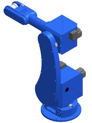
Fig 1. Robotic arm. (Credit: Prof. Jia-Yush Yen)
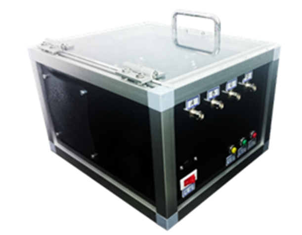
For promotion of stem cell proliferation/differentiation, we developed techniques and equipments based on ultrasound. Ultrasound can deliver mechanical waves into cells, inducing responses inside the cells. By using our ultrasound equipments (Figure 2), we are the first one to visualize the ultrasound-induced cellular responses such as lamellipodial (a typical response for cell activation) and mitosis.
Fig 2. Ultrasound equipment for cell culuture. (Credit: Prof. Jaw-Lin Wang)
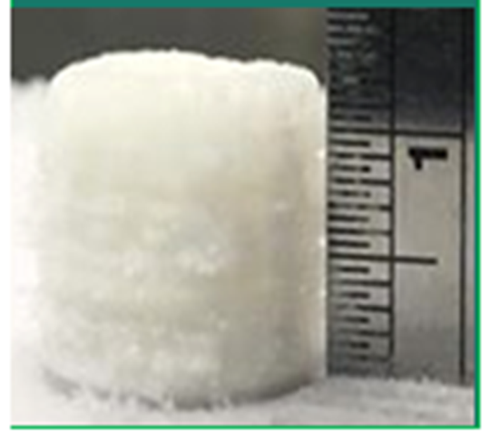
After preparation of enough stem cells with high quality, we then need stent to enclose these stem cells. Therefore, we developed bio-degradable stent material, which is suitable for human transplantation. This stent is multi-functional: its fine structure with patent allows the growth of stem cells and enclosure of small molecule drugs, thereby promoting regeneration of tracheal tissue. Moreover, out stent is 3D-printed, which allows completely customizable structure basing on medical imaging of individual patient (Figure 3).
These techniques and equipments have technological transfers and patents. In addition, animal study has been actively proceeded and clinical study will be started soon as the proof of concept of engineered bionic trachea.
Fig 3. 3D-printed tracheal stent. (Credit: Prof. Shan-Hui Hsu)
RELATED

STAY CONNECTED. SUBSCRIBE TO OUR NEWSLETTER.
Add your information below to receive daily updates.