

Mobile 5G enables smart living and digital economic development
Author(s)
Chia-Wei SunBiography
Chia-Wei Sun is currently an Associate Professor of the Department of Photonics, National Chiao Tung University, Hsinchu, Taiwan. His current research foci are diffuse optical tomography (DOT), near-infrared spectroscopy (NIRS), optical coherence tomography (OCT), neurophotonics and clinical applications based on biomedical optical imaging techniques.
Academy/University/Organization
National Chiao Tung University-
TAGS
-
Share this article
You are free to share this article under the Attribution 4.0 International license
- ENGINEERING & TECHNOLOGIES
- Text & Image
- January 21,2020
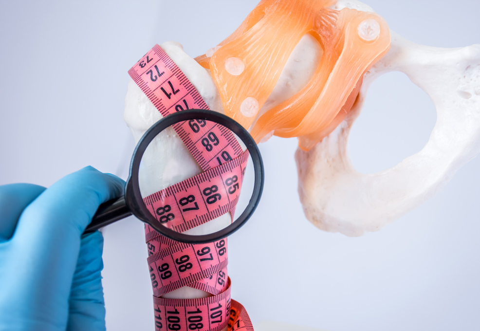
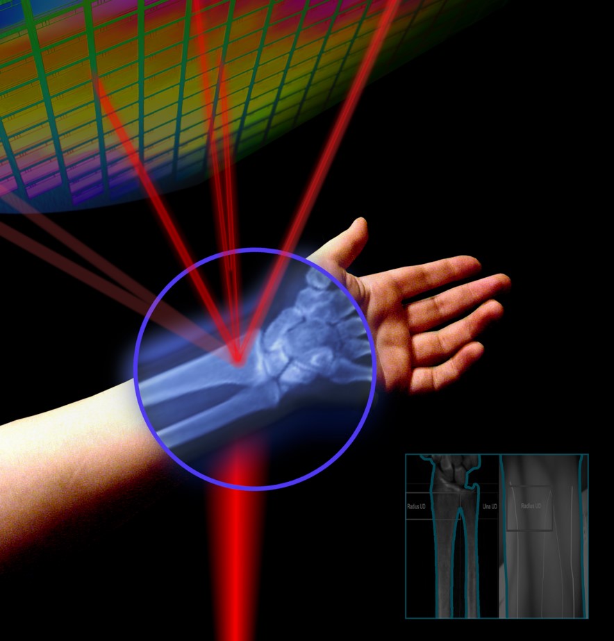
Osteoporosis, which is a disease associated with increased bone resorption and weakness, results in increasing risk of fractures among vertebrae in the spine, distal forearm and hip. However, the number of patients affected by osteoporosis is expected to increase with the growing geriatric population. Nowadays, osteoporotic fractures have become a major public health problem worldwide because of their associated morbidity, mortality and costs according to reports from the World Health Organization. About 20% of all patients suffering from hip fractures related to osteoporosis die within one year owing to complications resulting from being bedridden. Moreover, the number of days of hospitalization accounted for by hip fractures is even similar to that for cardiovascular disease, breast cancer and chronic obstructive pulmonary disease. Individuals suffering from osteoporotic-related fractures also consistently report significant reductions in quality-adjusted life years. Nevertheless, osteoporosis is a preventable and treatable disease. With prompt antiresorptive therapy, the osteoporosis-related fracture risk can be significantly reduced. Without a doubt, early detection and timely intervention are important to successfully manage osteoporosis and its associated complications. We provide an optical method to detect human distal radius bone density based on near infrared (NIR) imaging. The information of bone mineral density can be measured by the transluminational optical bone densitometric system. Compared to the dual-energy x-ray absorptiometry (DXA) results in clinical trials, NIR images show a strong correlation to DXA.
來源:https://doi.org/10.1002/jbio.201870148
Osteoporosis, which is a disease associated with increased bone resorption and weakness, results in increasing risk of fractures among vertebrae in the spine, distal forearm and hip. However, the number of patients affected by osteoporosis is expected to increase with the growing geriatric population. Nowadays, osteoporotic fractures have become a major public health problem worldwide because of their associated morbidity, mortality and costs according to the reports from the World Health Organization. About 20% of all patients suffering from hip fractures related to osteoporosis die within one year owing to complications resulting from being bedridden. Moreover, the number of days of hospitalization accounted for by hip fractures is even similar to that for cardiovascular disease, breast cancer and chronic obstructive pulmonary disease. Individuals suffering from osteoporotic-related fractures also consistently report significant reductions in quality-adjusted life years. Nevertheless, osteoporosis is a preventable and treatable disease. With prompt antiresorptive therapy, the osteoporosis-related fracture risk can be significantly reduced. Without a doubt, early detection and timely intervention are important to successfully manage osteoporosis and its associated complications.
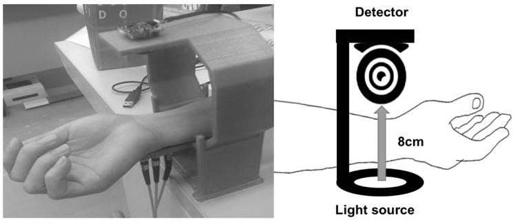
In our study, we offer an optical method to detect human distal radius bone density based on near infrared (NIR) imaging. The information of bone mineral density can be measured by the transluminational optical bone densitometric system. A simple optical device for measurement of distal radius bone density using the continuous wave (CW) NIR imaging system was designed in our experiment. Theoretically, the light intensity is attenuated after passing through the dense bone tissue. We measured the remnant light intensity (digit number, DN) after the NIR laser penetrated the distal forearm to clarify the association with the BMD of the distal radius. All participants received uniform examination with the distal forearm steadily located in the instrument (as shown in the figure below). The illuminating center of laser light focuses on the lister tubercle of the distal radius (UD part), which was selected in order to minimize the disturbance of light penetration ability because of less thickness of soft tissue coverage. The experimental result of our research demonstrated that the light intensity after the NIR laser penetrated the distal forearm was strongly and negatively correlated with the BMD measured with DXA at the UD part of the distal radius. The above-mentioned results indicate that the optical attenuation of our developed NIR image system could reflect the bone density of the distal radius to a great extent. Compared to the dual-energy x-ray absorptiometry (DXA) results in clinical trials, the NIR images show a strong correlation to DXA. To the best of our knowledge, our study is the first in vivo experiment to directly and simply demonstrate that the light intensity of NIR laser after bone penetration is strongly and negatively correlated with BMD. The result is expected to initiate the development of an optical bone density screening device to predict osteoporosis and future fracture risk.
RELATED


STAY CONNECTED. SUBSCRIBE TO OUR NEWSLETTER.
Add your information below to receive daily updates.