

A New World of Smart Healthcare

Author(s)
Shiuan-Yeh ChenBiography
Professor Shiuan-Yeh Chen received his BS/MS from the Department of Physics at National Taiwan University and his PhD from Duke University. He is currently associate Professor in the Department of Photonics at National Cheng Kung University. His research interests include: Raman tags and their biomedical application, Raman optical systems and strong coupling between light and emitters.
Academy/University/Organization
National Taiwan UniversitySource
https://www.osapublishing.org/oe/abstract.cfm?uri=oe-25-20-24767
https://www.osapublishing.org/boe/abstract.cfm?uri=boe-9-5-2142-
TAGS
-
Share this article
You are free to share this article under the Attribution 4.0 International license
- ENGINEERING & TECHNOLOGIES
- Text & Image
- January 21,2020
The median survival of the most fatal brain tumor, Glioblastoma (GBM) is 13 months. The complete resection of GBM is hindered by the infiltrative nature of GBM. Therefore, a robust and bright contrast agent will improve the image-guided surgery of GBM. In this report, the Raman tags based on core-satellite assemblies (CSA) are introduced and applied to GBM cell labeling. The Raman tags with non-resonant Raman reporters show higher stability than fluorophore-based Raman tags and free fluorophores. Because the Raman tags are selectively attached to the GBM cells instead of normal cells through EGFR antibodies, the contrast of the Raman image between GBM and normal cells is around 5-15. This Raman-based method can be integrated into the current image guide surgery to improve effectiveness of the GBM resection.
Glioblastoma (GBM), a fatal brain tumor, is difficult to eliminate by surgery due to its infiltrative nature. The median survival of GBM patient is around 13 months. Therefore, to enhance the contrast between GBM and normal cells, a robust image contrast agent for image-guided surgery is desired. Recently, the FDA approved ALA HCl (aminolevulinic acid hydrochloride) for the fluorescence image guided surgery of GBM; the derivative of ALA HCL (Protoporphyrin IX) generates fluorescence. The fluorophore-based images usually suffer from photo-bleaching, which degrades the image quality and precision of the surgery. In this article, Raman-based tags are introduced and applied to GBM cell labeling.
Fabrication and stability of Raman tags[1]: The backbone of the Raman tags is based on core-satellite assemblies (CSA) which consist of 50 nm and 20 nm gold nanoparticles. After the Raman reporter molecules are linked to the surface of core particles, the polyelectrolyte with positive charge coats 50 nm core particles to attract 20 nm satellite particles. Finally, the CSA are packaged with SiO2. The gap between the core and satellite particles is around 1 nm. The field amplitude of this gap field can be enhanced by the order of 200, which leads to strong Raman signals from Raman reporters within the gaps, Fig. 1.
![Fig. 1. Fabrication of the Raman tags. (Left) TEM image and (Right) Simulated field of the Raman tags [1].](/images/202001/0701.jpg)
Fig. 1. Fabrication of the Raman tags. (Left) TEM image and (Right) Simulated field of the Raman tags [1].
The three Raman tags with respective Raman reporters (Cy5, BDT, and DMcT) are tested and compared with the fluorescence of free Cy5 molecules (free fluorophores), Fig. 2(A). As shown in Fig. 2(B) during the 10 min illumination, Raman Tag2 and Tag3 show high stability. The Raman signals from Tag2 (658.9 cm−1 band) and Tag3 (1566.4 cm−1 band) have only a 10% intensity fluctuation. However, for the Raman Tag1, because part of the signal resulting from the resonant Raman effect decays due to the photobleaching of Cy5, the total Raman signal intensity of Raman Tag1 rapidly decreases to 18% in the first minute and then slowly decreases to ~5% after 5 min of illumination. For the free Cy5, the fluorescence immediately decays to <1% in the first minute. Therefore, the Raman tags embedded with non-fluorescent reporters (DMcT, BDT) show much higher stability than with fluorophore-based Raman reporters (Cy5) and free Cy5.
![Fig. 2. (A) From left to right: dark-field images, Raman images and the corresponding Raman spectra of three Raman tags. (B) The stability test of the Raman tags and free fluorophores [1].](/images/202001/0702.jpg)
Fig. 2. (A) From left to right: dark-field images, Raman images and the corresponding Raman spectra of three Raman tags. (B) The stability test of the Raman tags and free fluorophores [1].
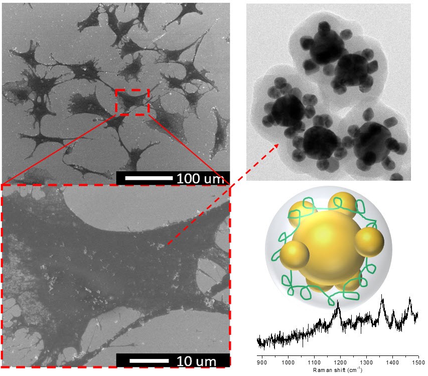
Image contrast of Raman tags labeled cells[2]: To quantitatively evaluate the imaging intensity contrast between the labeled GBM and normal cells, the GBM cells in the broadband Raman images and normal cells in the autofluorescence images are delineated first by the clear outlines obtained from the corresponding bright-field images. Then, the intensity within the cell outlines is integrated, divided by the area of the cells and then subtracted by the average background intensity in order to obtain the average Raman intensity, IGBM, or average autofluorescence intensity, Inormal. Under the same excitation conditions, for the cells labeled with intrinsic Raman tags, the tumor-normal ratio (TNR), IGBM/Inormal is ~5 while for using Cy5 embedded Raman tags, the TNR is ~15. In addition, the image collection time for cells labeled with intrinsic Raman tags and Cy5-embedded Raman tags is 10 s and 0.1 s, respectively. These results indicate that the Raman imaging based on these robust and brilliant tags has potential for real-time imaging, Fig. 3.
![Fig. 3. SEM image, broadband Raman image, and corresponding spectrum of a (A) GBM cell and (B) normal cell [2].](/images/202001/0703.jpg)
Fig. 3. SEM image, broadband Raman image, and corresponding spectrum of a (A) GBM cell and (B) normal cell [2].
Conclusion: The robust Raman tags provide high stability and brightness as a contrast agent for GBM cell labeling. The images of the labeled cells show clear contrast. These properties enable the tags to be integrated into the image-guided surgery for GBM.
Reference:
[1] Y.-C. Chang, L.-C. Huang, S.-Y. Chuang, W.-L. Sun, T.-H. Lin, S.-Y. Chen*, "Polyelectrolyte induced controlled assemblies for the backbone of robust and brilliant Raman tags", Optics Express, 25, 20, 24767-24779 (2017).
[2] L.-C. Huang, Y.-C. Chang, Y.-S. Wu, W.-L. Sun, C.-C. Liu, C.-I. Sze, S.-Y. Chen*, "Glioblastoma cells labeled by robust Raman tags for enhancing imaging contrast", Biomedical Optics Express, 9, 5, 2142-2153 (2018).
RELATED


STAY CONNECTED. SUBSCRIBE TO OUR NEWSLETTER.
Add your information below to receive daily updates.