

Computational Materials Design – From Materials Genome to the Development of New Materials and Their Applications

Author(s)
Meng-Tsan TsaiBiography
Meng-Tsan Tsai is a professor in the Department of Electrical Engineering at Chang Gung University in Taiwan. Dr. Tsai is also a joint appointment research fellow in the Department of Neurosurgery at Chang Gung Memorial Hospital in Taiwan. His research interests include optical imaging, laser surgery, and biomedical engineering.
Academy/University/Organization
Chang Gung Memorial Hospital-
TAGS
-
Share this article
You are free to share this article under the Attribution 4.0 International license
- ENGINEERING & TECHNOLOGIES
- Text & Image
- January 21,2020
Laser surgery has been a novel solution for various clinical applications such as ophthalmology and dermatology. In clinics, pulsed lasers become the key light sources to provide high energies for tissue ablation or heating. However, various clinical applications are limited by the extremely high cost of pulsed lasers. Additionally, determination of the treatment region is a key issue for laser surgery. To achieve precise treatment, we developed an integration system by combining laser surgery and optical coherence tomography for the diagnosis and treatment of diseases. Since the beam size of the treatment laser ranges from tens to hundreds of micrometers, optical coherence tomography could be a potential tool for the guidance of laser surgery due to its micrometer-scale resolution. Based on the scattering property of biological tissue, a three-dimensional microstructure can be acquired with optical coherence tomography without extra contrast agents. Such microstructures can be used to determine the lesion region. Aside from the acquisition of tissue microstructure, an imaging algorithm for three-dimensional angiography is demonstrated. With the guidance of optical coherence imaging, the abnormal tissue can be identified, and the laser treatment can be noninvasively monitored. The integrated system can effectively improve treatment accuracy and simultaneously evaluate the treatment outcome. Currently, the proposed technology has been successfully demonstrated for the airway, oral cavity, brain tumors and skin tumors.
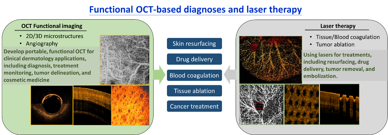
Laser surgery is an alternative solution for clinical treatment due to the advantages of less bleeding, reducing infections, and shorter recovery periods. Currently, laser surgery has been successfully applied in various applications including ophthalmology, dermatology, the gastrointestinal tract, the oral cavity including soft and hard tissues, and the spine. With laser surgery, tissue can be ablated or removed by being exposed to high optical energy. Moreover, the laser can be used for blood coagulation, which could be considered as a treatment strategy for diseases. Currently, pulsed lasers are the most popular for clinical applications because they can provide an extremely high peak energy to damage the targeted tissue without causing extra damage to the surrounding tissue. However, such short pulse lasers are typically bulky and very expensive. Additionally, the spot size of the treatment laser ranges from tens to hundreds of micrometers which can be used for treating a specific and small area. Limited to the spot size of the treatment laser, guidance with the imaging techniques will be necessary for accurate positioning and irradiation. Different imaging methods have been proposed as the guidance tools of laser surgery such as magnetic resonance imaging (MRI), computed tomography (CT), ultrasound imaging, microscopes, and endoscopes. However, since the imaging resolutions of MRI, CT, and ultrasound imaging are more than one hundred micrometers, it is difficult to identify a specific lesion for the laser surgery. Furthermore, although microscopes and endoscopes can achieve micrometer-scale resolutions, only the superficial layer of tissue can be visualized; therefore, it is difficult to investigate the thermal effect induced by the treatment laser in the deeper tissue layer.
In our study, we aimed to develop a theranostic system combining laser surgery and optical coherence tomography (OCT). The OCT technique is similar to ultrasound imaging, but uses light waves to probe the tissue structure instead of acoustic waves. According to the backscattered intensity from various depths of the biological tissue, the three-dimensional microstructure of the tissue can be acquired. Moreover, we have developed the algorithm of label-free, OCT-based angiography (OCTA) to investigate the tissue microcirculation. Typically, the OCT resolution of our systems can reach 3-10 m in biological tissue. With OCT/OCTA, normal and abnormal tissues can be differentiated, and then, the treatment laser can be accurately positioned. Moreover, the treatment results can be in vivo monitored with OCT/OCTA. Our research topics on the development of the OCT-guided laser surgery include the following issues.
- Develop a dual-regime fiber laser for OCT imaging and laser surgery: To reduce the complexity of the integrated system, a laser system with two output spectral regimes was developed including the 1.2 m regime for the OCT and the 1.55m regime for the laser surgery. Additionally, to easily combine the light source and the OCT system, the dual-regime laser is based on an all-fiber configuration. Such a design is able to effectively reduce the system complexity and cost.
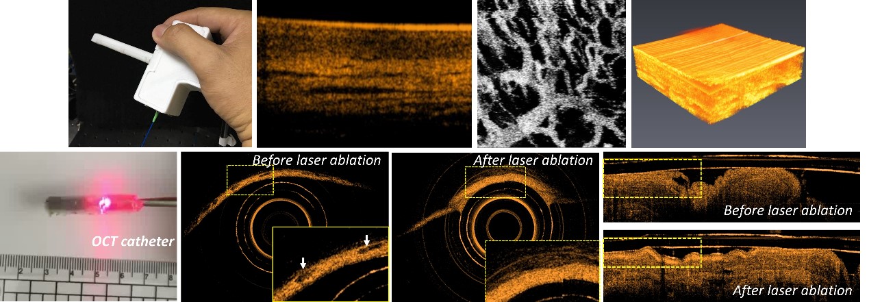
- Probe and catheter designs: In order to apply the developed OCT-guided laser surgery system to various applications, different scanning ends have been designed. In the past, the handheld probe for skin scanning was developed. For applications in the oral cavity, another handheld probe has been fabricated and the optical design of the oral probe enables scanning of the arbitrary sites of oral mucosae. All the developed probes can be used for the acquisition of the three-dimensional OCT/OCTA images and laser surgery. Additionally, optical catheters have been developed for scanning the inner organs such as the esophagus and gastrointestinal tract, while laser treatment can be simultaneously performed according to the OCT/OCTA results.
- Biological applications: In our studies, the developed system is used for applications in brain tumors, skin tumors, the oral cavity, and the gastrointestinal tract. For early-stage tumors, OCT is implemented to identify the tumor region, and the treatment laser can be accurately guided to the tumor center. Then, the treatment process can be monitored with OCT/OCTA. Furthermore, the catheter-based theranostic system is utilized for the removal of colon polyps with OCT as guidance.
- Exploit low-cost light sources for laser surgery: As mentioned above, short pulse lasers are commonly used for treatment, but the cost is extremely high. In our study, the economic laser diodes in the visible range are also implemented, and the induced thermal effect can be monitored and controlled with the OCT/OCTA to avoid extra damage to the surrounding tissue. The results illustrate that such low-cost laser diodes combined with the OCT/OCTA can also accurately ablate the abnormal tissue without extra damage and thereby greatly reduce the system cost.
With our developed technique, we have successfully proven that the developed theranostic system can be promising and the treatment efficiency can be greatly improved.
RELATED
STAY CONNECTED. SUBSCRIBE TO OUR NEWSLETTER.
Add your information below to receive daily updates.