

Magician on the spectrum - Multi-antenna System for mobile phones
Author(s)
Jung-Hsien ChiangBiography
Dr. Jung-Hsien Chiang is a distinguished professor of the Department of Computer Science and Information Engineering at National Cheng Kung University, and Chair of NCKU Hospital IT Office. Dr. Chiang’s prior research in bioinformatics has focused on intelligent text mining. He has contributed new techniques for developing biomedical text mining models, including data analytics, pattern extraction and literature classification. This leads to implementation of software prototypes to assist biomedical researchers in rapidly extracting useful knowledge from a huge number of biomedical documents and to improve understanding of gene function annotation.
Academy/University/Organization
National Cheng Kung UniversitySource
https://focustaiwan.tw/sci-tech/202004130022-
TAGS
-
Share this article
You are free to share this article under the Attribution 4.0 International license
- ENGINEERING & TECHNOLOGIES
- Text & Image
- June 21,2020
As we continue to face the rapid increase in confirmed Coronavirus cases around the world, we created an AI-based pneumonia detection platform for COVID-19.The system is able to automatically detect high-risk patients with pneumonia which will then send information to doctors. With that information, doctors are able to make decisions and provide a treatment plan for the diagnosis. Any x-ray images taken for patients will be automatically uploaded to the system. Our system will process the images to assist doctors in determining whether the patients are infected. If the test result is positive, doctors will receive an e-Alert (on computer or mobile phone) that contains the original scans and detection results. Doctors from the Department of Medical Imaging provided us with thousands of positive and negative chest x-rays of pneumonia cases. Thus, our team created a UNet-based deep learning model to automatically detect the presence of pneumonia with this dataset. We consulted with engineers in order to better integrate our model to assist doctors with their diagnoses. Transfer of x-ray scans, server construction, and prediction response were all needed to build this e-Alert system. Our system has already been tested with and adopted by doctors at the National Cheng Kung University Hospital. The system achieved 92% accuracy in detecting the pneumonia symptoms, based on 1,400 medical images. To further assist precise diagnoses, we will develop the new approach and model to improve the performance of detection and enhance the ability to detect potential COVID-19 patients.
The COVID-19 pneumonia aggravates in the rapid progression stage (3-7 days after the onset of the initial symptoms) presenting bilateral multi-lobe light consolidations with air-bronchograms inside that could be detected early on chest x-rays. Therefore, an automatic chest X-ray pneumonia detection system was developed in the last two years with the aim of assisting radiologists in diagnosing pneumonia more quickly. The basic idea is to use an artificial intelligence (AI) system for quickly recognizing pulmonary infiltrates on chest x-ray images with an e-alert system that can detect pneumonia infection in lungs and automatically alert doctors more quickly.
Our research team created a UNet++ machine learning model that can detect the presence of pneumonia. Using this learning model, the system is able to automatically distinguish high-risk patients by scanning their chest x-rays to determine more quickly whether they are infected with pneumonia. If the x-ray image proves positive, the doctor will receive an e-alert, either via computer or mobile phone, containing both the original scans and the detection results.
In the beginning, our team used chest X-ray images published by Stanford University, which contain more than 60,000 patients’ data for the past 15 years, as our training data to develop the AI-based pneumonia detection model. To test the model robustness, we further used 3,000 pneumonia and non-pneumonia chest X-ray image data published by NIH. Unfortunately, it did not end well. After several intensive discussions with clinical doctors, we concluded that the major reason is that the quality of the images from NIH is uneven. Since the image data sources differ, the purpose of taking X-ray examinations and the corresponding labels are both various. This kind of problem was not discovered in the past, causing the limitation of the model application. We were finally able to build the AI system due to the team obtaining more than 1,000 chest x-rays from patients suspected of having pneumonia, provided by the NCKU Department of Medical Imaging. So far, the system has already been used at NCKU Hospital and has greatly increased diagnosis efficiency.
Based on the 1,400 images the system has managed to scan so far, it has achieved 92 percent accuracy in the detection of pneumonia symptoms.
For enhancing the ability of feature extraction on the model, we re-designed the model structure by using the squeeze-and-excitation function, which can extract more fine-grained features in the process. On the other hand, we utilized the Feature Pyramid Network to extend the receptive field on the convolutional kernel, which improved the performance of the pneumonia detection with various locations on the chest x-ray images. In the training phase, we applied the data augmentation, including flipping, rotation, adding noises, and image distortion, to make the model more robust and to avoid it overfitting.
With the coronavirus pandemic over the past six months, the system has also been used for early detection of potential COVID-19 patients, and it only takes a single second to determine if the chest X-ray scan of a patient needs further screening for coronavirus, as shown in the figures. Both cases were confirmed patients from China and the U.S.A., respectively.
In the future, the research team will include functions for other types of screening, such as CT scans or MRIs, to provide doctors with even more accurate diagnostic information for early detection of confirmed patients.
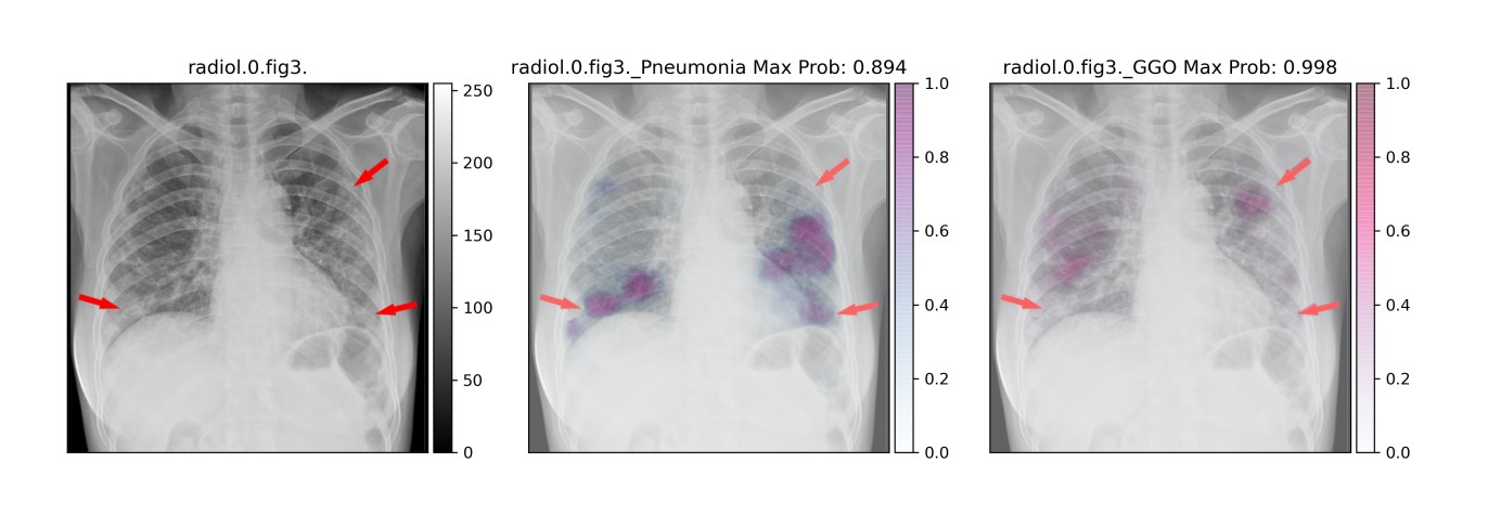
Example of confirmed patient from Wuhan, China showing the detection result with probability of 0.99
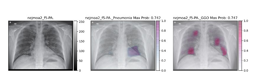
Example of confirmed patient from Seattle, U.S.A. showing the detection result with probability of 0.75
RELATED


STAY CONNECTED. SUBSCRIBE TO OUR NEWSLETTER.
Add your information below to receive daily updates.